Can mentalizing interacting shapes lead to empathy in humans?
How do you think empathy comes about in humans? Can simple shapes arouse empathy? How does it relate to autistic people?
We investigate following questions:
- Do people empathize with socially interacting shapes?
- Which networks in the brain are associated with empathy?
- Is there any kind of correlation among the regions involved in social cognition which leads to empathy?
- Is absence of a task or experimental stimuli (i.e resting state), good control for such tasks?
Brief scientific background
The perceptions of the participants can be directly attributed to the ability to reflect on the emotional and instinctive responses in others often referred to as ‘mentalizing’. However, one concept that goes hand in hand with mentalizing is empathy.
Method and Analysis
Dataset Used:
We used the The Human Connectome Project dataset. In the Social Cognition aspect of the Human Connectome Project dataset, the dataset consists of observations for 339 participants (called subjects). For each subject, BOLD signals are collected for resting state and active state. BOLD signals are observed due to the difference in magnetic properties of oxygenated and deoxygenated blood, which is captured by the MRI machine.
In the task based fMRI, the data was collected through two runs. In each run, 5 videos with mental and random interaction each of 23 seconds were shown to subjects with a fixation time of 15s between successive videos, and the response for each video from the participant was recorded. Each run had either 2 videos with mental interaction and 3 videos with random interaction or 3 videos with mental interaction and 2 videos with random interactions. The response from participants recorded belonged to one of the three categories: Mental Interaction, Random Movement, and Not sure.
Analysis 1: Subtraction Analysis
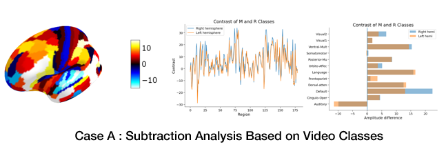
- Each comparision contains analysis of three aspects:
- Contrasts in Parcels for both hemispheres
- Contrasts in network activations for both hemispheres
- Visualizing contrasts of activitations in parcels
- 4 different comparisons
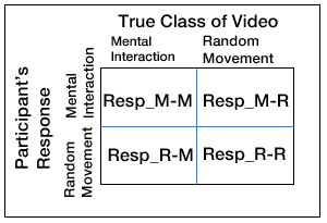
This is the nomenclature that we follow for response of participants and condition of videos:
- Case A. Mental and Random Classes of Videos
- Case B. Resp_M-M and Resp_R-R (Taking Resp_R-R Sate as Control)
- Case C. Resp_M-M and Resting State (Taking Resting Sate as Control)
- Case D. Resp_M-M and Res_R-M
Analysis 2: Correlation Analysis for Functional Connectivity
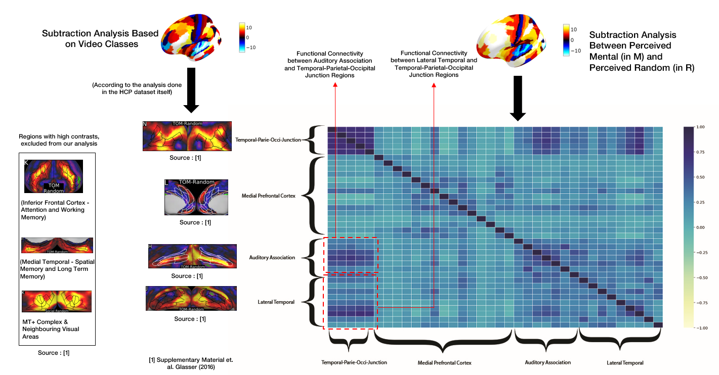
We use correlation analysis to investigate the functional connectivity among the regions responsible for empathy. We investigated 36 parcels, consistent with the literature, belonging to the following four regions: Temporal-Parietal-Occipital Junction, Medial prefrontal cortex,Auditory Association, and Lateral Temporal cortex We arranged these parcels regions-wise in the correlation matrix.
As you can see in the figure (Rightmost), Temporal-parietal-occipital junction was found to be correlating well with both auditory association and Lateral temporal region, hinting that their functional connectivity leads to empathy.
(Middle Column) These are contrasts in the activations in the four regions, that were observed when subtraction analysis was performed on the basis of only the class of videos.
(Leftmost Column) There were also three other regions where we observed high activations, dedicated for cognition tasks other than empathy, and hence we excluded them from our analysis, namely : Inferior Frontal Cortex, which is responsible for Attention and Working Memory, Medial Temporal cortex, responsible for Spatial Memory and Long Term Memory, MT+ Complex & Neighbouring Visual Areas which are responsible for visual processing.
Conclusion
We observed:
-
Correlations between temporal-parietal-occipital junction, auditory association region, lateral temporal region, and medial-prefrontal cortex, manifesting the underlying connectivity of social cognition.
-
More clear contrast when the true random was considered as control as compared to the resting state condition.
-
Subtraction analysis yielded better results when the participant responses were considered as compared to when only condition-based analysis is performed.
-
The subtraction analysis between the perceived mental and random interactions for the mental condition resulted in relatively high contrast in a region close to the posterior superior temporal sulcus. This is the same region found to be less active for the people having Autism.
It was an amazing experience from figuring out the research question, to diving into the dataset, reframing the hypothesis as and when we gain new insights and performing the analysis.
Back to the Future
-
The observed finding can be extended to explain the clinical disorders by doing the same experiment with people having autism where it’s assumed that there is reduced social cognition or people having schizophrenia, where there can be increased social cognition, due to visualizing what actually is not there and perceiving an interaction with it.
-
And as we now know, Mentalizing, empathy and mirroring of emotional response are closely related. We’d also try to further investigate if we can comment on the presence of mirror neurons.


